• Skip to primary navigation
  • Skip to main content
  • Skip to primary sidebar
  • Skip to footer

  (514) 667-9541

  REQUEST APPOINTMENT

HOME

ABOUT

DR PIZEM

FRANÇAIS

Endomontreal Root Canal Clinic logo

ENDOMONTREAL

Dr Pizem Advanced Endodontics Clinic

  • Advanced Root Canal Treatment
    • Symptoms & Procedure
    • Why Micro Endodontics?
  • General Dentistry
  • Patients info
    • Frequently asked questions (FAQ)
    • Need a root canal revision?
    • Treatment sequence at our clinic
    • Tooth saving tips
  • For Dentists
    • Refer a patient
    • Clinical cases
    • Implants vs Endodontics
Contact Us

Carl Zeiss Opmi Proergo Microscope VS Complete Stenosis of an Apical Root Canal Split

You are here: Home / Atypical canal configurations / Carl Zeiss Opmi Proergo Microscope VS Complete Stenosis of an Apical Root Canal Split

March 22, 2011 By Dr Pierre Pizem Leave a Comment

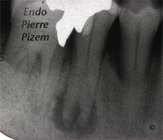
Atypical Canal Configuration, Type V, Root Canal Treatment Pre Therapy 48744-1
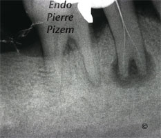
Atypical Canal Configuration, Type V, Root Canal Treatment Per Therapy 48744-1
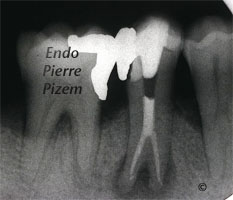
Atypical Canal Configuration, Type V, Root Canal Treatment Per Therapy 48744-2
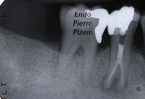
Atypical Canal Configuration, Type V, Root Canal Treatment Post Therapy 48744-1
Atypical Canal Configuration, Type V, Root Canal Treatment Post Therapy 48744-2

Calcified Canals. Case Study Number 487445

Clinical examination: Sinus tract, mobility: 0, deciduous amalgam restoration.

Radiographic examination: Alveolar bone with circumscribed lucency, apical root canal split, hypertaurodontism (bull’s tooth), apical root canal split branches not visible on X ray dental film, hypercementosis

Diagnosis: pulpal necrosis with chronic periapical infection,

Etiology: marginal leakage, caries

Root canal procedure:

First appointment: gaining access to the split, locating entries, shaping and cleaning apical root canal branches inserting intracanal medication for 8 days.

Second appointment: intracanal medication retrieval, copious CHX 2% irrigation, drying canals and permanent root canal obturation with Pulp Canal Sealer and gutta percha (lateral and vertical condensation).

In that specific case, microscope was most helpful during all the following steps necessary to insure a better prognosis for this patient:

1) Locating and gaining access to buccal and lingual root canal entries (apical split was clearly visible under magnified observation)

2) Striving to find a third branch in apical split minimizing the chances of omitting an untreated canal

3) Aiming at the right root canal orifice when:

  • Inserting the two first endodontic files to confirm canal lengths
  • inserting a file sequence to shape and clean each canal
  • Positioning irrigating syringe needle tip and calcium hydroxide syringe tip toward the right canal entry
  • Inserting absorbent paper points in both canals when drying canals
  • Inserting master, accessory gutta percha cones and finger plugger when doing final obturation with lateral and vertical condensation

4) Checking for pulpal tissue remnants prior to final obturation to minimise the chances of pushing them back into the apical area

Category iconAtypical canal configurations,  D.O.M. versus completely calcified systems,  Dental operating microscope (D.O.M.) assisted R.C.T.,  Extreme endo clinical cases,  NEW CASES,  Taurodontism,  Type V Tag iconabsorbent paper points,  apical delta,  bull tooth,  calcified canals,  Calcium hydroxide,  complete root canal stenosis,  Dental expertise,  Dental operative microscope,  dental radiograph,  Dystrophic calcifications,  Endodontic expertise,  endodontic file,  Endodontics,  Gutta percha,  hypercementosis,  hypertaurodontism,  image,  microendodontics,  Pain,  periapical radiograph,  radiolucent apical lesion,  root canal apical split,  root canal procedure,  Root canal system calcifications,  Sinus tract,  vertucci type V root canal configuration

Reader Interactions

Leave a Reply Cancel reply

Your email address will not be published. Required fields are marked *

Primary Sidebar

Quality Endodontics

 30+ Years of Experience
in Root Canal Treatment

 300+ Clinical Cases
Published for dentists

 Conveniently Located
Near Metro Vendôme

Call for an appointment:

(514) 667-9541

Cases with high level of difficulty:

Pierre Pizem Endodontics

A little message from Dr Pizem:

« I understand how stressful going to the dentist could be for you, the patient. So let me reassure you. I’ve built my entire reputation on my professionalism and the passion I pour into my craft. I will not compromise on equipment quality, nor on the effort necessary to restore your teeth. In the end, my greatest satisfaction is to see you fully recover your dental health, so you may grab big bites at life again. »

Ready to start your treatment?

In order to avoid risks of further complications, we recommend you book a visit as early as you can. However, if you're already feeling pain or urgent symptoms, we'll make every attempt to see you today.

Get in touch

Footer

Contact

(514) 667-9541

REQUEST APPOINTMENT

Monday
8:30 am - 4:30 pm
Tuesday
8:30 am - 4:30 pm
Wednesday
8:30 am - 4:30 pm
Thursday
8:30 am - 4:30 pm
Friday
Closed
Sat. Sun.
Closed

Clinic info

  • Home
  • About
  • Dr Pizem
  • Publications
  • Referring Dentists
    • Refer a patient
    • Clinical cases
  • Français

Patient info

  • Root Canal Therapy
  • General Dentistry
  • Endodontic Retreatment
  • FAQ New Patients
  • Tooth Saving Tips
5025 Sherbrooke St. W, Suite 330A
Montreal (QC) H4A 1S9
Near Metro Vendôme       Directions

Copyright © 2025 Endomontreal

Return to top