• Skip to primary navigation
  • Skip to main content
  • Skip to primary sidebar
  • Skip to footer

  (514) 667-9541

  REQUEST APPOINTMENT

HOME

ABOUT

DR PIZEM

FRANÇAIS

Endomontreal Root Canal Clinic logo

ENDOMONTREAL

Dr Pizem Advanced Endodontics Clinic

  • Advanced Root Canal Treatment
    • Symptoms & Procedure
    • Why Micro Endodontics?
  • General Dentistry
  • Patients info
    • Frequently asked questions (FAQ)
    • Need a root canal revision?
    • Treatment sequence at our clinic
    • Tooth saving tips
  • For Dentists
    • Refer a patient
    • Clinical cases
    • Implants vs Endodontics
Contact Us

OPMI PROergo Dental Operative Microscope VS Completely Calcified Canals

You are here: Home / Dental operating microscope (D.O.M.) assisted R.C.T. / D.O.M. versus completely calcified systems / OPMI PROergo Dental Operative Microscope VS Completely Calcified Canals

May 4, 2011 By Dr Pierre Pizem Leave a Comment

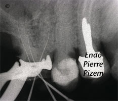
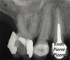
Dental operating microscope (D.O.M.), D.O.M. versus completely calcified systems, Root Canal Treatment Pre-Therapy 465016-1Dental operating microscope (D.O.M.), D.O.M. versus completely calcified systems, Root Canal Treatment Per-Therapy 465016-1Dental operating microscope (D.O.M.), D.O.M. versus completely calcified systems, Root Canal Treatment Per-Therapy 465016-2Dental operating microscope (D.O.M.), D.O.M. versus completely calcified systems, Root Canal Treatment Post-Therapy 465016-1

Case Study Number 465016

Patient has been referred to to us in order to complete a previously started root canal treatment on completely calcified root canal system. Pulp chamber is obliterated with embedded pulpstones, root canals are not visible on preoperative X ray dental film, these are calcified canals.

Patient is given full knowledge of the possible risks and benefits of such a complex procedure.  Patient is in pain and she wants to keep her own tooth and give an informed consent.

Dystrophic calcifications have been removed from pulp chamber with ultrasonic diamond coated tips. Operative field observation is enhanced with high magnification and coaxial xenon lamp illumination. Once located root canal entries had to be widened in order to progressively regain patency in each root canal. Second X ray shows our first instruments in the root canals, these are K Files 06. Canals have then been shaped to K file size 15 and calcium hydroxide inserted. Following appointment allowed us to finish shaping, cleaning and obturation of the root canal system. Final outcome can be seen on fourth X ray dental film.

Category iconD.O.M. versus completely calcified systems,  Dental operating microscope (D.O.M.) assisted R.C.T.,  Pulp stones (denticles),  Root canal systems calcifications Tag iconAdherent pulpstones,  calcified canals,  complete root canal stenosis,  Dental operative microscope,  Denticles,  Dystrophic calcifications,  Embedded pulpstones,  Mb2,  microendodontics,  root canal procedure,  Root canal treatment

Reader Interactions

Leave a Reply Cancel reply

Your email address will not be published. Required fields are marked *

Primary Sidebar

Quality Endodontics

 30+ Years of Experience
in Root Canal Treatment

 300+ Clinical Cases
Published for dentists

 Conveniently Located
Near Metro Vendôme

Call for an appointment:

(514) 667-9541

Cases with high level of difficulty:

Pierre Pizem Endodontics

A little message from Dr Pizem:

« I understand how stressful going to the dentist could be for you, the patient. So let me reassure you. I’ve built my entire reputation on my professionalism and the passion I pour into my craft. I will not compromise on equipment quality, nor on the effort necessary to restore your teeth. In the end, my greatest satisfaction is to see you fully recover your dental health, so you may grab big bites at life again. »

Ready to start your treatment?

In order to avoid risks of further complications, we recommend you book a visit as early as you can. However, if you're already feeling pain or urgent symptoms, we'll make every attempt to see you today.

Get in touch

Footer

Contact

(514) 667-9541

REQUEST APPOINTMENT

Monday
8:30 am - 4:30 pm
Tuesday
8:30 am - 4:30 pm
Wednesday
8:30 am - 4:30 pm
Thursday
8:30 am - 4:30 pm
Friday
Closed
Sat. Sun.
Closed

Clinic info

  • Home
  • About
  • Dr Pizem
  • Publications
  • Referring Dentists
    • Refer a patient
    • Clinical cases
  • Français

Patient info

  • Root Canal Therapy
  • General Dentistry
  • Endodontic Retreatment
  • FAQ New Patients
  • Tooth Saving Tips
5025 Sherbrooke St. W, Suite 330A
Montreal (QC) H4A 1S9
Near Metro Vendôme       Directions

Copyright © 2025 Endomontreal

Return to top